Understanding the Dermatome Map of the Leg: A Complete Information
Associated Articles: Understanding the Dermatome Map of the Leg: A Complete Information
Introduction
With nice pleasure, we’ll discover the intriguing matter associated to Understanding the Dermatome Map of the Leg: A Complete Information. Let’s weave attention-grabbing data and provide recent views to the readers.
Desk of Content material
Understanding the Dermatome Map of the Leg: A Complete Information
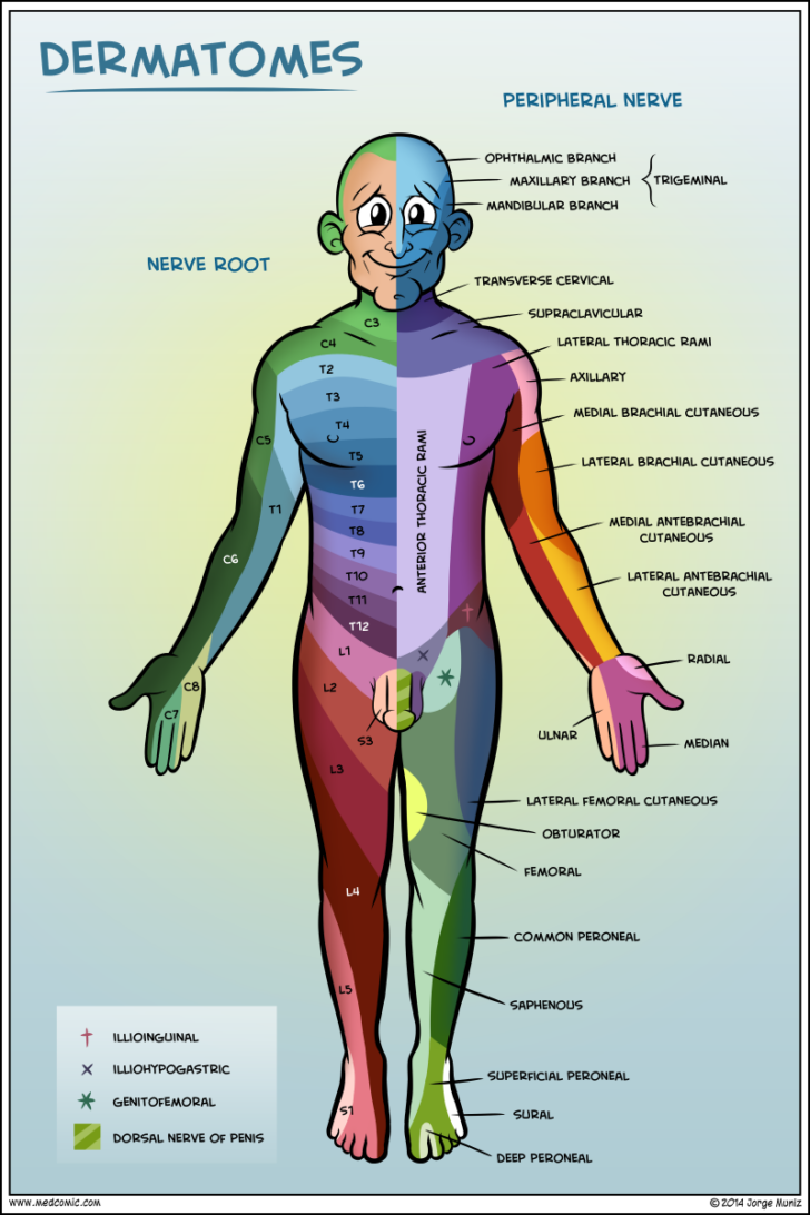
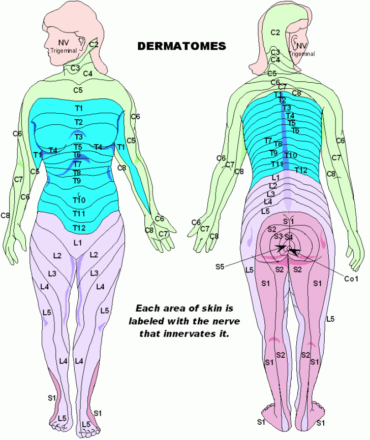
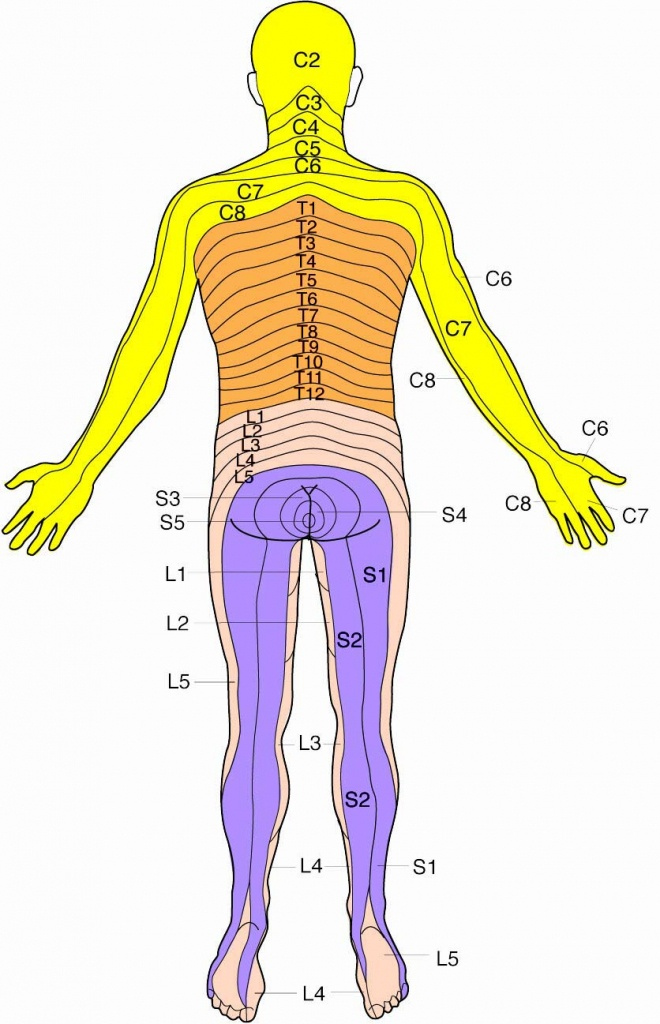
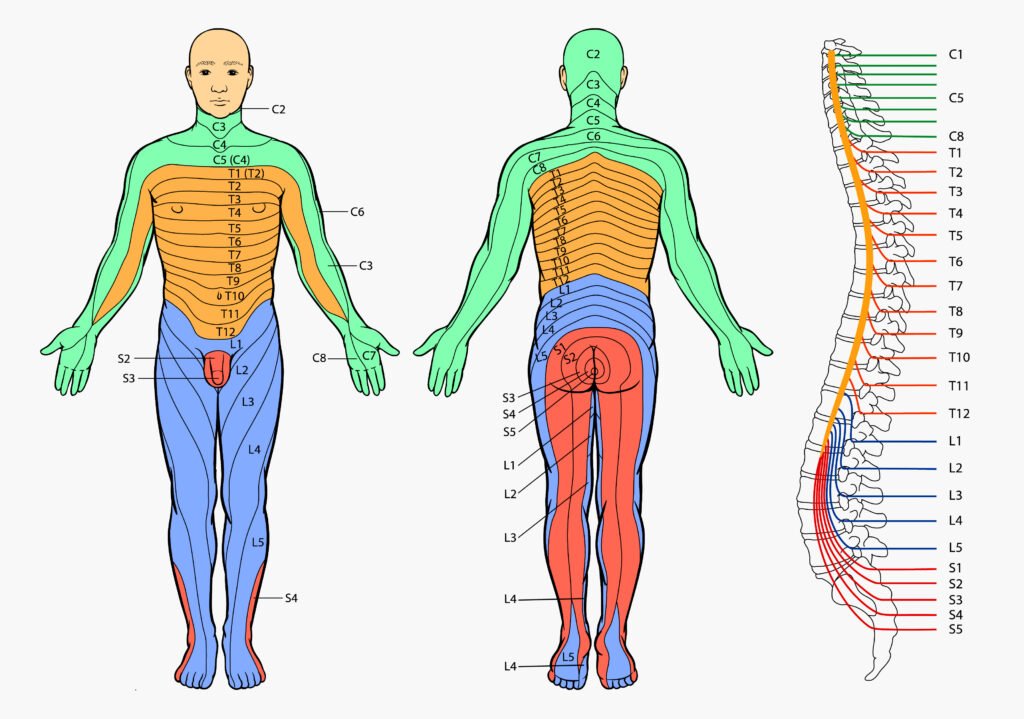
The human physique’s intricate nervous system is liable for transmitting sensory data from the periphery to the central nervous system (CNS) and motor instructions from the CNS to muscular tissues. A key part of this method is the dermatome, a particular space of pores and skin innervated by a single spinal nerve. Mapping these dermatomes gives essential data for diagnosing neurological situations affecting the spinal twine and peripheral nerves. This text will delve into the dermatome map of the leg, exploring its construction, medical significance, and implications for analysis and remedy.
What are Dermatomes?
Dermatomes are areas of pores and skin which might be primarily equipped by sensory fibers from a single posterior spinal root. Every spinal nerve, shaped by the union of dorsal (sensory) and ventral (motor) roots, emerges from the spinal twine at a particular degree. The sensory fibers inside these nerves carry details about contact, temperature, ache, and strain from a particular space of pores and skin again to the spinal twine. This space of pores and skin is the dermatome. Whereas there’s important overlap between adjoining dermatomes, making exact delineation difficult, a basic map may be constructed. This map is invaluable in medical follow for localizing neurological lesions.
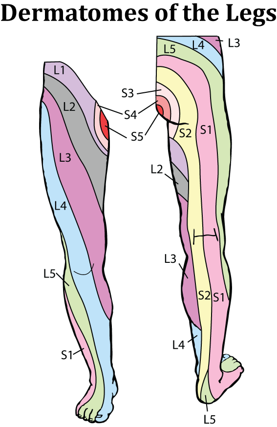
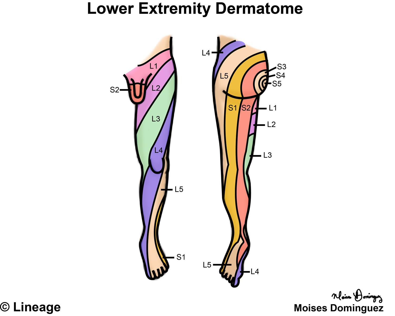
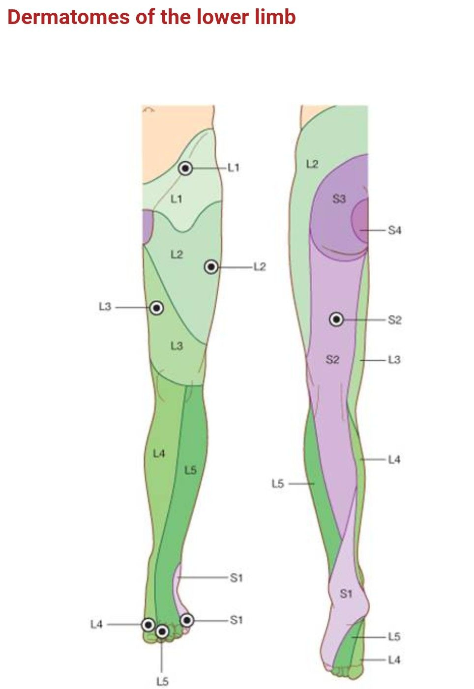
The Leg’s Dermatomal Group:
The leg’s dermatomal innervation originates from the lumbar (L1-L5), sacral (S1-S5), and coccygeal (Co1) spinal nerves. These nerves department out to produce the assorted areas of the leg, together with the anterior thigh, medial thigh, posterior thigh, lateral leg, medial leg, and foot. The distribution isn’t completely segmental, with appreciable overlap and variation between people. Nevertheless, a basic sample is persistently noticed:
-
L1: A small space of the anterior groin and higher medial thigh. The contribution of L1 is usually minimal and may be troublesome to exactly outline.
-
L2: Covers a bigger space of the anterior and medial thigh, extending barely down the medial side of the knee.
-
L3: Continues down the medial thigh, encompassing the medial knee and a portion of the medial leg.
-
L4: Innervates the medial side of the leg, extending to the medial malleolus (ankle bone) and the medial side of the foot, together with the large toe.
-
L5: Provides the lateral side of the leg, extending to the dorsum (high) of the foot, together with the primary and second toes.
-
S1: Innervates the lateral side of the foot and the only real, together with the little toe.
-
S2-S4: These dermatomes primarily provide the posterior thigh, posterior leg, and the only real of the foot. S2 is mostly thought of to cowl the higher posterior thigh, S3 the center, and S4 the decrease.
-
S5: Contributes to the lateral side of the foot and the only real.
-
Co1: Innervates a small space of the pores and skin over the coccyx.
Medical Significance of the Leg Dermatome Map:
The dermatome map is a vital software for neurologists and different healthcare professionals in diagnosing a variety of neurological situations. By fastidiously assessing the sensory distribution of signs, clinicians can pinpoint the seemingly degree of spinal twine or nerve root involvement. For instance:
-
Radiculopathy: This situation includes compression or irritation of a spinal nerve root, leading to ache, numbness, tingling, or weak spot within the corresponding dermatome. A affected person experiencing ache radiating down the lateral leg and the dorsum of the foot would possibly counsel L5 radiculopathy.
-
Spinal Wire Lesions: Injury to the spinal twine at a particular degree will end in sensory loss within the corresponding dermatomes under the extent of the lesion. The sample of sensory loss may help decide the extent and placement of the spinal twine damage.
-
Peripheral Neuropathy: This situation includes harm to peripheral nerves, which may trigger sensory disturbances in varied dermatomes. The sample of sensory loss may help establish the precise nerves affected.
-
Shingles (Herpes Zoster): This viral an infection impacts the sensory ganglia of a single dermatome, leading to a attribute painful rash confined to that dermatome. The dermatome map is crucial in diagnosing and managing shingles.
-
Surgical Procedures: Data of dermatomes is significant throughout surgical procedures involving the backbone or peripheral nerves to attenuate harm to sensory nerves and to foretell potential postoperative sensory adjustments.
Limitations of Dermatome Maps:
Whereas dermatome maps are clinically priceless, it is essential to acknowledge their limitations:
-
Particular person Variation: The precise distribution of dermatomes can differ considerably between people. Anatomical variations and particular person variations in nerve branching patterns can result in inconsistencies.
-
Overlap: Important overlap exists between adjoining dermatomes, making it difficult to pinpoint the precise degree of involvement in some instances.
-
Referred Ache: Ache may be skilled in areas exterior the affected dermatome, complicating the interpretation of sensory findings.
-
Incomplete Sensory Loss: Not all sensory fibers from a single spinal nerve could also be affected in a lesion, leading to incomplete sensory loss throughout the dermatome.
Assessing Dermatomes in Medical Observe:
Clinicians make the most of varied strategies to evaluate dermatomes and establish potential neurological points:
-
Sensory Testing: This includes utilizing a pinprick, gentle contact, or temperature stimulus to evaluate the affected person’s sensation in several areas of the leg. The clinician compares the affected person’s sensation to the anticipated dermatomal distribution.
-
Reflex Testing: Deep tendon reflexes (e.g., patellar, Achilles) are assessed to judge the integrity of the corresponding spinal nerve roots and their motor operate.
-
Muscle Power Testing: Muscle power is assessed to judge the motor operate of the muscular tissues innervated by the spinal nerves.
-
Imaging Research: Imaging strategies reminiscent of MRI and CT scans can present detailed anatomical details about the spinal twine and nerves, confirming the medical findings from sensory testing.
Conclusion:
The dermatome map of the leg is a priceless software for understanding the sensory innervation of the decrease limb. Its medical purposes are intensive, aiding within the analysis and administration of assorted neurological situations. Whereas particular person variation and overlap exist, cautious medical evaluation, mixed with different diagnostic instruments, permits clinicians to precisely interpret the sensory findings and formulate acceptable remedy plans. Understanding the dermatome map is crucial for any healthcare skilled concerned within the evaluation and administration of sufferers with neurological issues affecting the decrease limb. Nevertheless, it is essential to do not forget that the map serves as a information, and medical judgment and a complete neurological examination are important for correct analysis. Additional analysis continues to refine our understanding of dermatomal distribution and its medical implications.

Closure
Thus, we hope this text has offered priceless insights into Understanding the Dermatome Map of the Leg: A Complete Information. We thanks for taking the time to learn this text. See you in our subsequent article!